A Diagram Of Joints And Bones In The Human Body : Cartilage is an extremely flexible type of tissue, which is why it is located around joints.. The skeleton of the human body is made out of bones and the cartilage supporting those bones. Human skeleton, the internal skeleton that serves as a framework for the body. Elbow joint diagram human elbow elbow joint elbow bones the diagram depicts bones and parts of a human elbow including humerus, trochiea, ulna, radius, head, neck, radial fossa and others. At a synchondrosis, the bones are united by hyaline cartilage. The ulna is one of two bones that give structure to the forearm.
Hinge joints allow movement in one direction, as seen in the knees and elbows. Human skeleton anatomy human anatomy drawing human anatomy and physiology anatomy study anatomy reference pose reference anatomy bones heart anatomy body anatomy. A joint, or articulation, is the junction between two or more bones. In humans and other vertebrates, the bones form a framework called the skeletal system that provides structure and shape. At a synchondrosis, the bones are united by hyaline cartilage.
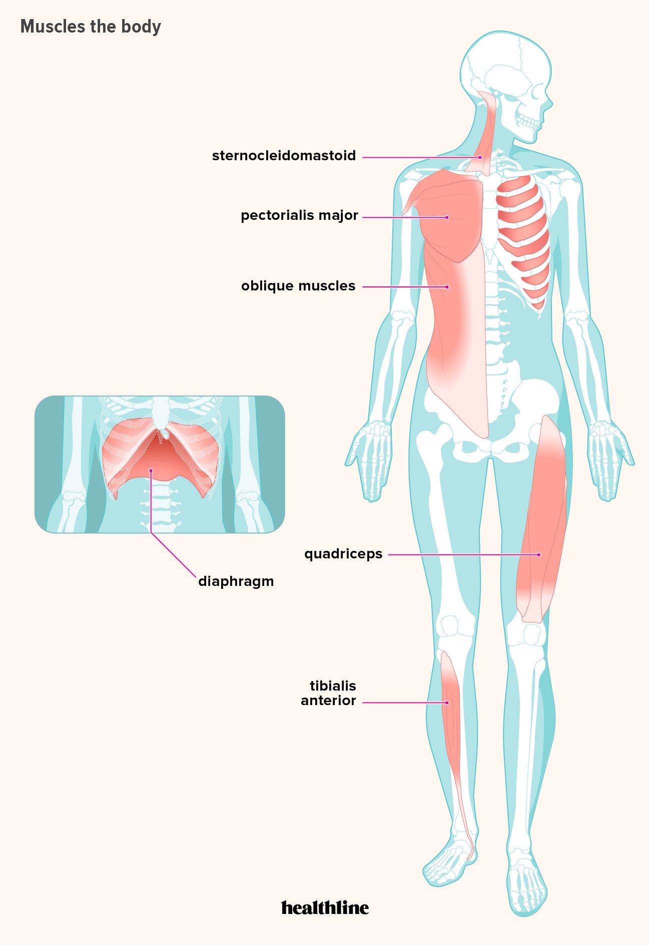
How Many Muscles Are In The Human Body Plus A Diagram from post.healthline.com Did you ever play the game operation as a kid? If bones provide the framework, the joints provide the flexibility by permitting movement. Muscles and bones of the human body 12 photos of the muscles and bones of the human body anatomy bones of the human body quiz, major muscles and bones in the human body, muscles and bones in the human body, number of muscles and bones in the human body, the muscles. Yet the hip joint is also one of our most flexible joints and allows a greater range of motion than all other joints in the body except for the shoulder. Did you take biology in school? Identify and explain the 3 types of joint structures found in the human body. The ulna is located on the opposite side of the forearm from the thumb. Joints hold the skeleton together and support movement.
The first is by joint function, also referred to as range of motion.the second way to categorize joints is by the material that holds the bones of the joints together;
That is an organization of joints by structure. It joins with the humerus on its larger end to make the. But, you may be surprised to hear that most of the bones are in the feet and hands. Elbow joint diagram human elbow elbow joint elbow bones the diagram depicts bones and parts of a human elbow including humerus, trochiea, ulna, radius, head, neck, radial fossa and others. There are three main types: At a symphysis, the bones are joined by fibrocartilage. All of these joints are synovial joints. The six types of synovial joints allow the body to move in a variety of ways. A joint is an area where 2 or more bones are in contact with each other. The bones of the pelvis are the hip bones, sacrum, and coccyx. The human body has over 200 bones. There also are bands of fibrous connective tissue—the ligaments and the tendons—in intimate relationship with the parts of the skeleton. They hold up your body, and along with your muscles, keep you moving.
The hips and shoulders have this type of joint, in which the round end of a long bone fits into the hollow. Synovial joints are subdivided based on the shapes of the articulating surfaces of the bones that form each joint. There are three main types: A joint is an area where 2 or more bones are in contact with each other. The ulna is located on the opposite side of the forearm from the thumb.
Muscular And Skeletal Systems from www2.estrellamountain.edu For instance, the human foot has 26 bones each. Yet the hip joint is also one of our most flexible joints and allows a greater range of motion than all other joints in the body except for the shoulder. Human skeleton, the internal skeleton that serves as a framework for the body. That is an organization of joints by structure. Bones of the hand diagram, find out more about bones of the hand diagram. Joints are points where a muscle is. Did you take biology in school? This framework consists of many individual bones and cartilages.
There are two ways to categorize joints.
Cross section of human bone diagram 12 photos of the cross section of human bone diagram cross section diagram of human bone, bone, cross section diagram of human bone. Without your bones, you'd just be one big blob! Joints hold the skeleton together and support movement. The muscle acts as the effort force; The long bones of the body contain many distinct regions due to the way in which they develop. An example of a pivot joint is the joint between the first two vertebrae in the spine. This can lead to many health issues such as bone thinning, kidney damage, heart problems and even death. Synovial joints are subdivided based on the shapes of the articulating surfaces of the bones that form each joint. At a synchondrosis, the bones are united by hyaline cartilage. Identify and explain the 3 types of joint structures found in the human body. Identify and explain the 3 range of motions found in human joints. Main joints of the body. The following activities will help your students learn how the bones, muscles, and joints work together, as well as how to prevent injuries from occurring.
Bones of the hand diagram, find out more about bones of the hand diagram. The major key factors that help in locomotion are bones and muscles. Either way, that may sound like a lot of bones. Each hip bone contains three bones— the ilium, ischium, and pubis — that fuse together as we grow older.the sacrum, five fused. The bones of the leg are the femur, tibia, fibula and patella.the foot bones shown in this diagram are the talus, navicular, cuneiform, cuboid, metatarsals and calcaneus.
Anatomy Of The Human Shoulder Joint from www.verywellhealth.com Other sesamoid bones can form in the joints of the hands and feet, but are not present in all people. The muscle acts as the effort force; Human skeleton, the internal skeleton that serves as a framework for the body. This diagram shows the six classes of movable joints in the human body. Joints are points where a muscle is. A joint, or articulation, is the junction between two or more bones. K to grade 2 • human body series bones, muscles, and joints. Human skeleton anatomy human anatomy drawing human anatomy and physiology anatomy study anatomy reference pose reference anatomy bones heart anatomy body anatomy.
They hold up your body, and along with your muscles, keep you moving.
There are two ways to categorize joints. There also are bands of fibrous connective tissue—the ligaments and the tendons—in intimate relationship with the parts of the skeleton. Joints in the human skeleton can be grouped by function (range of motion) and by. Human skeleton anatomy human anatomy drawing human anatomy and physiology anatomy study anatomy reference pose reference anatomy bones heart anatomy body anatomy. Compare the structure of joints with their range of motions. Yet the hip joint is also one of our most flexible joints and allows a greater range of motion than all other joints in the body except for the shoulder. Synovial joints are subdivided based on the shapes of the articulating surfaces of the bones that form each joint. For instance, the human foot has 26 bones each. Human skeleton labeled human skeleton bones human skeleton anatomy human body anatomy human anatomy and physiology anatomy of the body skeleton figure skeleton model human skull. 10+ mesmerizing learn to draw people the female body ideas. It allows us to walk, run, and jump. This can lead to many health issues such as bone thinning, kidney damage, heart problems and even death. The human body has over 200 bones.


/shoulder_pain_medreview-01-5c3b9f8546e0fb0001bdeaaa-d0a4923b7a3d441fb12d992c454a8ca7.png)


0 Komentar